Home

מקום כורסה סיור fluorescent microscopy markers אנציקלופדיה כניסה נקבוביות

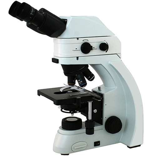
Fein Optic RB30 UV / DAPI Fluorescence Microscope

Improved Chemical-Genetic Fluorescent Markers for Live Cell Microscopy |  Biochemistry
Improved Chemical-Genetic Fluorescent Markers for Live Cell Microscopy | Biochemistry

Improved Plasmids for Fluorescent Protein Tagging of Microtubules in  Saccharomyces cerevisiae - Markus - 2015 - Traffic - Wiley Online Library
Improved Plasmids for Fluorescent Protein Tagging of Microtubules in Saccharomyces cerevisiae - Markus - 2015 - Traffic - Wiley Online Library

Fluorescence markers for advanced microscopy From photophysics to biology –  2024 – March 17-22 2024, Les Houches, France
Fluorescence markers for advanced microscopy From photophysics to biology – 2024 – March 17-22 2024, Les Houches, France
Solved Fluorescence microscopy using antibodies tagged with | Chegg.com

Live mitochondria seen in unprecedented detail: photobleaching in STED  microscopy overcome
Live mitochondria seen in unprecedented detail: photobleaching in STED microscopy overcome

Ecole des Houches: Fluorescence markers for advanced microscopy -  France-BioImaging
Ecole des Houches: Fluorescence markers for advanced microscopy - France-BioImaging

Blog - Keeping it Simple with Fluorescence

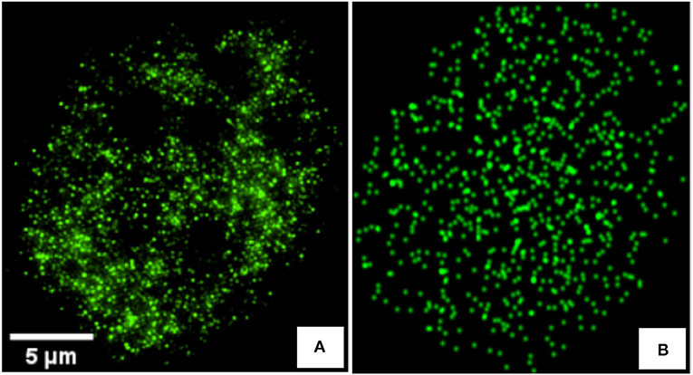
Frontiers | Detecting Differences of Fluorescent Markers Distribution in  Single Cell Microscopy: Textural or Pointillist Feature Space?
Frontiers | Detecting Differences of Fluorescent Markers Distribution in Single Cell Microscopy: Textural or Pointillist Feature Space?

Colocalization of fluorescent markers in confocal microscope images of  plant cells | Nature Protocols
Colocalization of fluorescent markers in confocal microscope images of plant cells | Nature Protocols
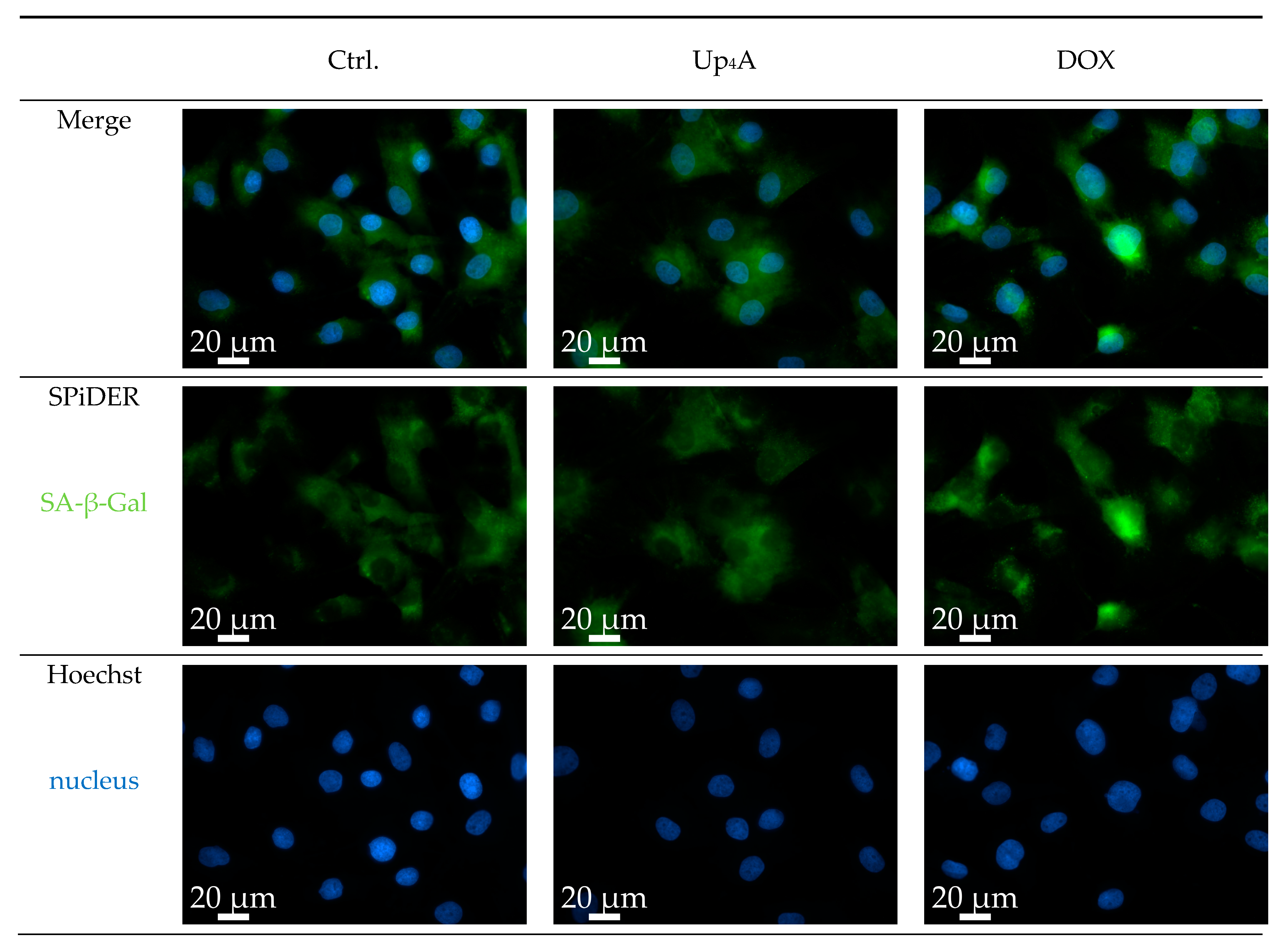
IJMS | Free Full-Text | A Novel Protocol for Detection of Senescence and  Calcification Markers by Fluorescence Microscopy
IJMS | Free Full-Text | A Novel Protocol for Detection of Senescence and Calcification Markers by Fluorescence Microscopy
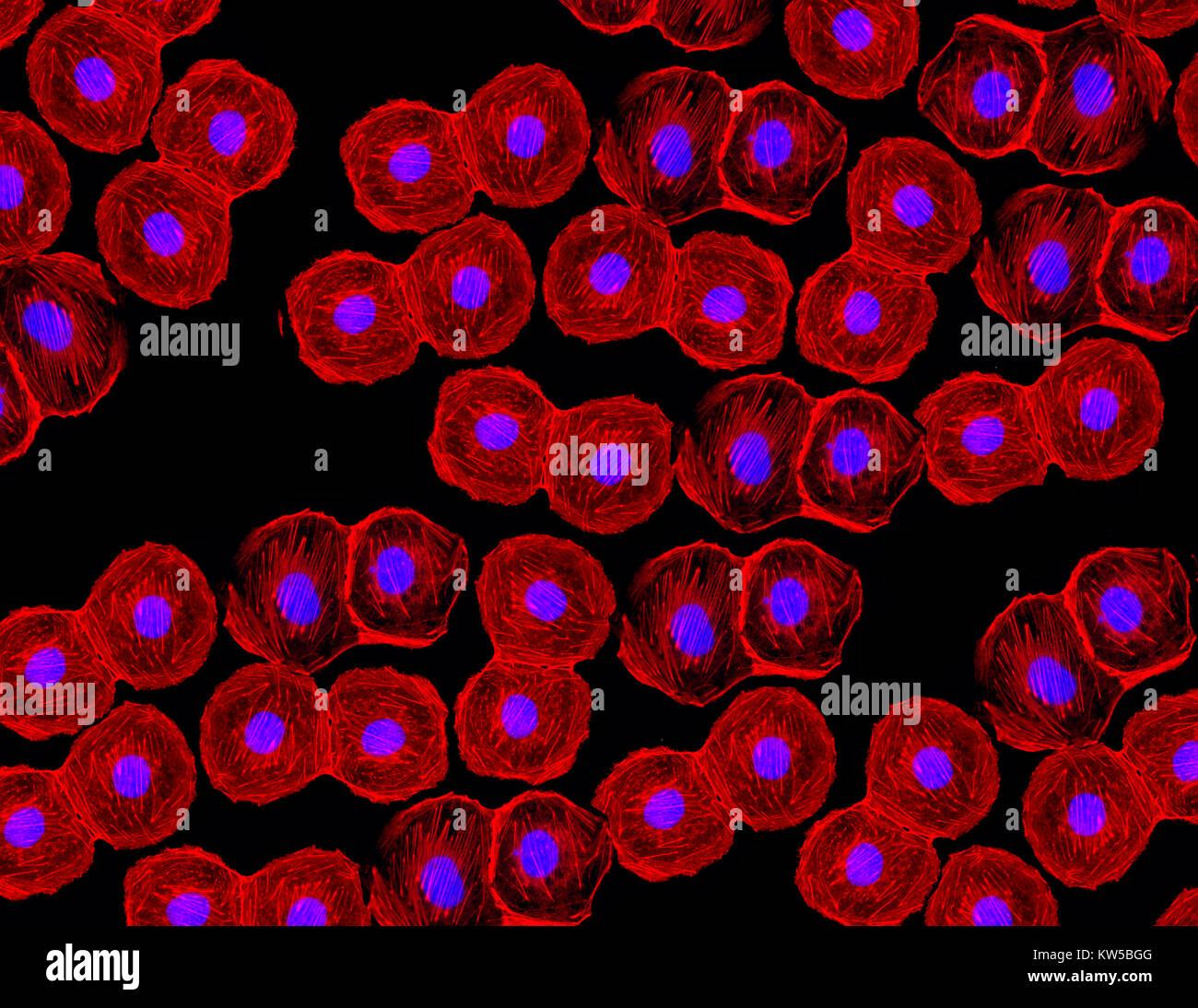
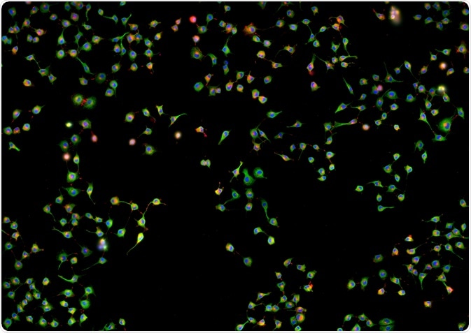
Fluorescent image of human stem cells stained with monoclonal antibodies  markers under the microscopy showing nuclei in blue and microtubules in red  Stock Photo - Alamy
Fluorescent image of human stem cells stained with monoclonal antibodies markers under the microscopy showing nuclei in blue and microtubules in red Stock Photo - Alamy

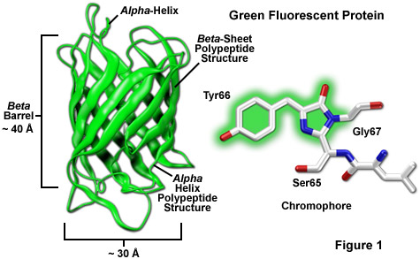
ZEISS Microscopy Online Campus | Introduction to Fluorescent Proteins

Utilizing Uncertainty Estimation in Deep Learning Segmentation of Fluorescence  Microscopy Images with Missing Markers | DeepAI
Utilizing Uncertainty Estimation in Deep Learning Segmentation of Fluorescence Microscopy Images with Missing Markers | DeepAI

A) Fluorescence microscopy images of the expression of the... | Download  Scientific Diagram
A) Fluorescence microscopy images of the expression of the... | Download Scientific Diagram

Importance of Colocalization Studies

Fluorescence microscope - Wikipedia

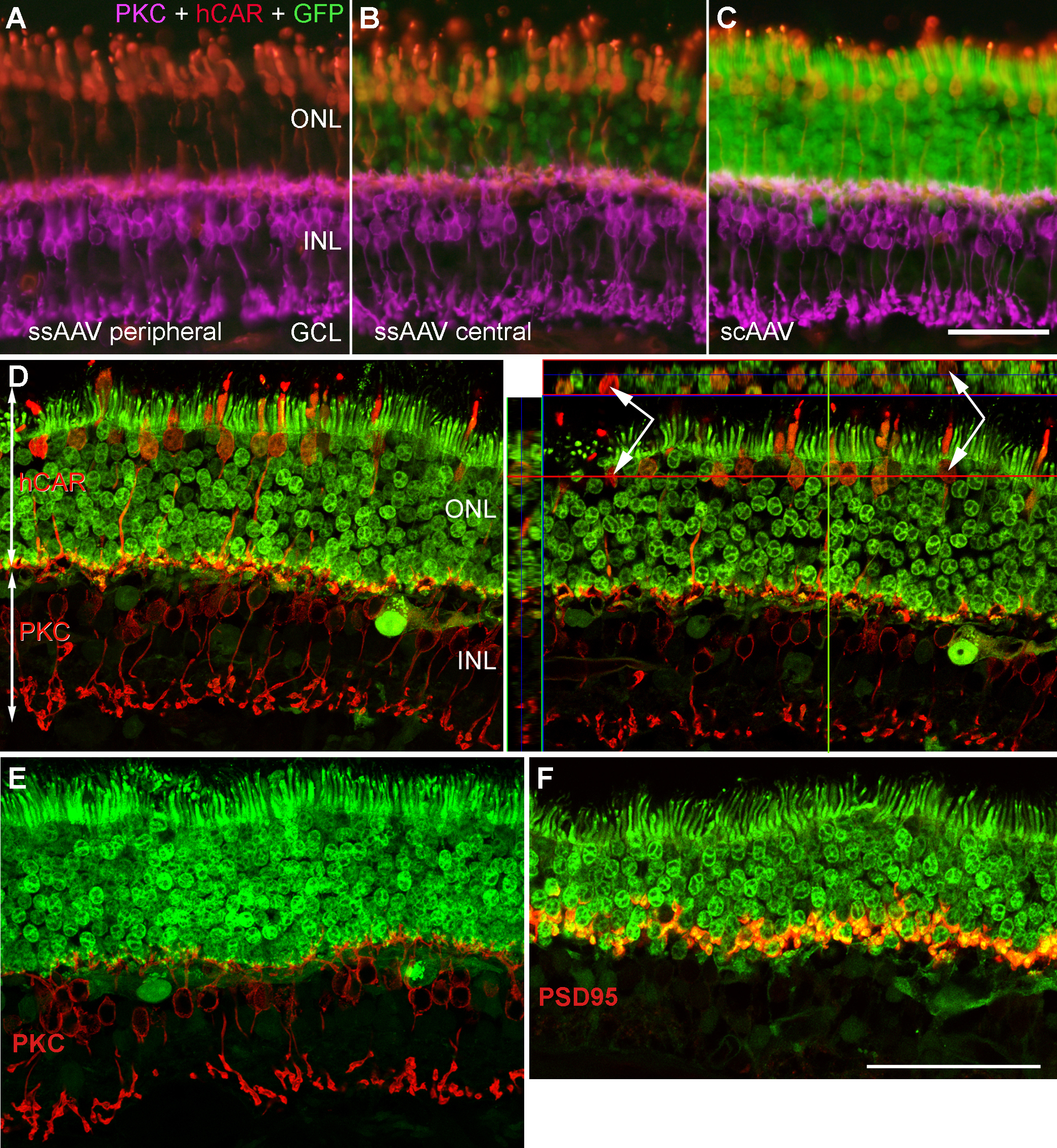
Molecular Vision: Petersen-Jones, Mol Vis 2009; 15:1835-1842. Figure 2

Fundamentals of Fluorescence Microscopy: Exploring Life with Light |  SpringerLink
Fundamentals of Fluorescence Microscopy: Exploring Life with Light | SpringerLink

Fluorescein diacetate 5-maleimide, Fluorescent marker used in microscopy  studies (CAS 150322-01-3) (ab145333)
Fluorescein diacetate 5-maleimide, Fluorescent marker used in microscopy studies (CAS 150322-01-3) (ab145333)

Introduction to the Quantitative Analysis of Two-Dimensional Fluorescence  Microscopy Images for Cell-Based Screening | PLOS Computational Biology
Introduction to the Quantitative Analysis of Two-Dimensional Fluorescence Microscopy Images for Cell-Based Screening | PLOS Computational Biology

VWO Masterclass Fluorescenctie Microscopie – Microscopycourse

2.3. Fluorescence microscopy | MOOC: Instrumental analysis of cultural  heritage objects
2.3. Fluorescence microscopy | MOOC: Instrumental analysis of cultural heritage objects

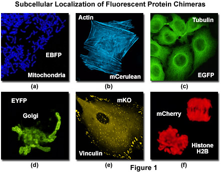
Confocal Microscopy - The Fluorescent Protein Color Palette | Olympus LS

A photostable fluorescent marker for the superresolution live imaging of  the dynamic structure of the mitochondrial cristae | PNAS
A photostable fluorescent marker for the superresolution live imaging of the dynamic structure of the mitochondrial cristae | PNAS

Quantitative and Dynamic Assessment of the Contribution of the ER to  Phagosome Formation - ScienceDirect
Quantitative and Dynamic Assessment of the Contribution of the ER to Phagosome Formation - ScienceDirect